
Make Room for Growth
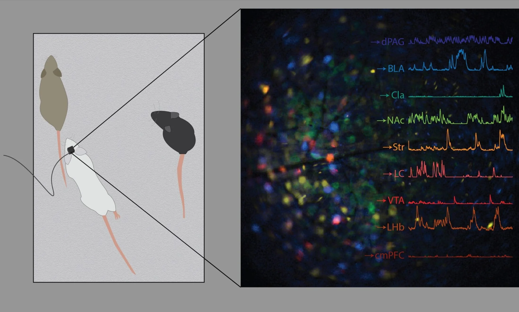
Neuroplex allows scientists to measure and link neural activity of up to 9 unique cell types in behaving animals, significantly accelerating data collection.
In Brief
Neuroplex, a novel imaging pipeline, enables simultaneous measurements of the activity of up to nine identified brain cell types in living mice
This approach accelerates insights into how neural circuits work together to control behavior
Scientists at the Max Planck Florida Institute for Neuroscience (MPFI), in collaboration with ZEISS and MetaCell, have developed a powerful new imaging pipeline called Neuroplex. Published in eLife, the technique allows simultaneous monitoring of the activity of up to nine distinct neuronal populations in freely moving mice, dramatically accelerating the pace of scientific exploration into how the brain controls behavior.


The Challenge
For years, neuroscientists linking brain activity to behavior have faced a fundamental limitation: miniscopes, the tiny head-mounted microscopes used to observe neural activity in behaving animals, could capture neural activity, but couldn’t reliably distinguish more than two different types of brain cells at a time.
“To understand the brain, we need to link patterns of activity in specific neurons to behavior,” stated lead author Dr. Mary Phillips. “We can readily use labels to color-code different populations of neurons, but when using miniscopes to correlate neural activity to behavior, we couldn’t distinguish more than two of these populations. This made it difficult to compare the activity across multiple cell types and circuits to understand how specific circuits regulate behavior.”
To work around this, researchers were forced to test one cell type at a time, repeating the same behavioral experiments, but labeling distinct neuron types each time. This iterative process, however, was slow and costly. It also prevented direct comparison of different neuron types within the same animal, muddying conclusions due to differences among individual animals. As an alternative, scientists delineated different neuron types after the behavioral experiment by removing and slicing brain tissue, color-coding different neuron types, then imaging the processed brain tissue using microscopes that can distinguish multiple colors. However, matching the cells imaged with a miniscope in a living animal to those in post-mortem, processed brain tissue was challenging and low-throughput, resulting in significant data loss. Additionally, this approach destroyed the ability to track the activity of identified cell types over time to determine how their activity changes with learning, aging, or during disease progression.
Related Articles

Quisque iaculis facilisis lacinia. Mauris euismod pellentesque tellus sit amet mollis.